
An In-Depth Exploration: In Vivo Morphology of the Human Cornea — Unraveling the Microscopic Landscape

The cornea, a transparent and dome-shaped structure at the front of the eye, plays a critical role in vision by providing approximately two-thirds of the eye's focusing power. Understanding the in vivo morphology of the cornea, the study of its microscopic structure and shape, is crucial for gaining insights into its physiological functions and the development of targeted therapeutic interventions. This article delves into the world of in vivo corneal morphology, exploring techniques, findings, and their implications in ophthalmology.
In Vivo Imaging Techniques for Corneal Morphology Assessment
Technological advancements have led to the development of sophisticated imaging modalities that enable non-invasive visualization and quantification of corneal morphology in vivo. Key techniques include:
5 out of 5
| Language | : | English |
| File size | : | 31067 KB |
| Text-to-Speech | : | Enabled |
| Screen Reader | : | Supported |
| Enhanced typesetting | : | Enabled |
| Print length | : | 110 pages |
1. Optical Coherence Tomography (OCT)
OCT utilizes low-coherence interferometry to generate cross-sectional images of the cornea with micron-scale resolution. It provides detailed information on corneal thickness, curvature, and the laminar structure of the various corneal layers.
2. Scheimpflug Imaging
Scheimpflug photography captures images of a rotating cornea, providing three-dimensional maps of its anterior and posterior surfaces. It allows for accurate measurements of curvature, elevation, and corneal asphericity.
3. Confocal Microscopy
Confocal microscopy uses a laser scanning mechanism to generate high-resolution images of the corneal epithelium and stroma. It enables visualization of cellular architecture, nerve fiber distribution, and other microstructural details.
Microscopic Anatomy of the Cornea
The cornea is composed of five distinct layers:
1. Epithelium
The epithelium, the outermost layer, is a non-keratinized, stratified squamous epithelium that provides protection and contributes to tear film formation.
2. Bowman's Layer
Bowman's layer is a thin, acellular layer of collagen fibrils that lies beneath the epithelium. It provides structural support and resistance to mechanical stress.
3. Stroma
The stroma, the thickest layer, is composed of densely packed, regularly arranged collagen fibrils that provide the cornea with its transparency and strength.
4. Descemet's Membrane
Descemet's membrane is a thin, elastic layer that separates the stroma from the endothelium. It plays a role in maintaining corneal hydration and transparency.
5. Endothelium
The endothelium is a single layer of polygonal cells that lines the posterior surface of the cornea. It regulates corneal hydration and ion transport, ensuring corneal transparency.
In Vivo Morphology Findings and Clinical Implications
In vivo morphological studies have provided valuable insights into the structural characteristics and variations of the human cornea. Key findings include:
1. Corneal Thickness and Curvature
OCT and Scheimpflug imaging have revealed variations in corneal thickness and curvature across the corneal surface, influencing refractive power and vision quality. Thinning of the central cornea is associated with keratoconus, a progressive corneal disease, while increased curvature is seen in astigmatism.
2. Stromal Architecture
Confocal microscopy has allowed researchers to examine the intricate arrangement of collagen fibrils in the corneal stroma. Disruption of the stromal architecture, such as in corneal scars or dystrophies, can lead to impaired corneal transparency and visual distortions.
3. Epithelial Morphology
In vivo imaging techniques have revealed variations in epithelial cell size, shape, and thickness. Alterations in epithelial morphology have been associated with dry eye syndrome and other ocular surface disorders.
4. Endothelial Morphology
The density and morphology of endothelial cells can be assessed using specular microscopy. Reduced endothelial cell count and changes in cell shape are indicators of endothelial dysfunction, which can result in corneal edema and vision impairment.
In vivo corneal morphology provides a wealth of information about the microscopic structure and shape of the human cornea. Advanced imaging techniques have enabled ophthalmologists to non-invasively assess corneal morphology, aiding in the diagnosis, monitoring, and management of various corneal disorders. Understanding the structural characteristics of the cornea is essential for developing targeted therapies that preserve and restore corneal function, ultimately improving vision outcomes.
Image Captions
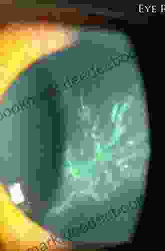
**Figure 1:** Cross-sectional OCT image of the human cornea. Different corneal layers are clearly distinguishable. **Figure 2:** Scheimpflug image showing the three-dimensional surface topography of the cornea. **Figure 3:** Confocal microscopic image revealing the regular arrangement of collagen fibrils in the corneal stroma.
5 out of 5
| Language | : | English |
| File size | : | 31067 KB |
| Text-to-Speech | : | Enabled |
| Screen Reader | : | Supported |
| Enhanced typesetting | : | Enabled |
| Print length | : | 110 pages |
Do you want to contribute by writing guest posts on this blog?
Please contact us and send us a resume of previous articles that you have written.
 Book
Book Story
Story Genre
Genre Reader
 Library
 E-book
 Paragraph
 Sentence
 Shelf
Shelf Glossary
 Preface
 Annotation
 Manuscript
 Scroll
 Codex
Codex Bestseller
 Library card
 Autobiography
 Memoir
 Encyclopedia
 Dictionary
 Character
 Librarian
 Card Catalog
 Borrowing
 Study
Study Scholarly
 Lending
 Reserve
 Academic
 Journals
 Reading Room
 Rare Books
 Special Collections
 Interlibrary
 Thesis
 Dissertation
 Reading List
 Book Club
 Textbooks
 Laurie Frankel
 Washington Irving
 Mario Pescatori
 Jacqueline L Jackson
 Tom Mcmillan
 Ed Sobey
 Emma King
 Lauren Castillo
 Bil Howard
 Gail Tuchman
 Elizabeth Mccallum Marlow
 Anne Ylvisaker
 Anonym
 Philip Duke
 Jo Ann Petrucci Andrews
 Richard A Nielsen
 Suzanne Goldring
 Jill Switzer
 Seth Zuiho Segall
 Tom Holladay
Light bulbAdvertise smarter! Our strategic ad space ensures maximum exposure. Reserve your spot today!

 Cristian CoxSleepless Nights: A Haunting Literary Journey Through the Dark Heart of New...


 Douglas FosterFollow ·14k
 Ed CooperFollow ·5k
 Terry PratchettFollow ·16.9k
 Allan JamesFollow ·2.6k
 Derek BellFollow ·7.1k
 Blake KennedyFollow ·2.4k
 Reed MitchellFollow ·8.3k
 Damon HayesFollow ·5.5k

 Oscar Wilde
Don't Stop Thinking About the Music: Exploring the Power...
Music is an...

 Floyd Richardson
Snowman Story Problems Math With Santa And Friends
It's a cold winter day, and...

 W. Somerset Maugham
What Every Classroom Teacher Needs To Know: A...
Teaching is a challenging...

 Edgar Cox
Tall Tales But True: A Lifetime of Motorcycling...
I've been riding motorcycles for over 50...

 Chinua Achebe
Buni: Happiness Is a State of Mind
Buni is a beautiful...

 Herman Melville
The Arts and Crafts of Older Spain: Embodying the Essence...
In the heart of the Iberian...
5 out of 5
| Language | : | English |
| File size | : | 31067 KB |
| Text-to-Speech | : | Enabled |
| Screen Reader | : | Supported |
| Enhanced typesetting | : | Enabled |
| Print length | : | 110 pages |
