
Diagnostic Procedures in Ophthalmology by H.V. Nema

Ophthalmology, the branch of medicine dealing with the diagnosis and treatment of eye disorders, relies heavily on a range of diagnostic procedures to assess eye health and identify potential issues. These procedures play a crucial role in guiding treatment decisions and ensuring optimal patient outcomes. This article provides a comprehensive overview of the most commonly used diagnostic procedures in ophthalmology, offering detailed descriptions and images to enhance understanding and provide valuable insights for ophthalmologists, optometrists, and other healthcare professionals.
4.5 out of 5
| Language | : | English |
| File size | : | 55845 KB |
| Print length | : | 462 pages |
| Screen Reader | : | Supported |
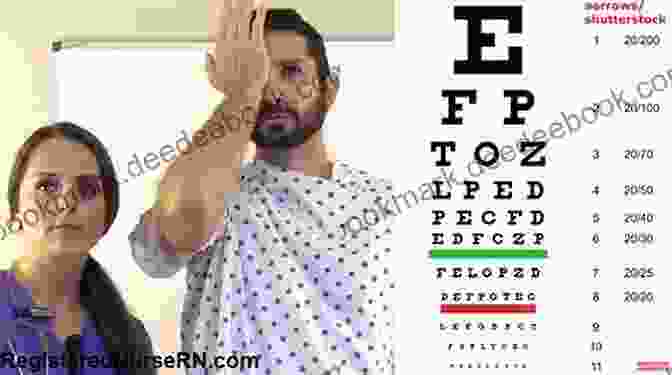
Visual Acuity Testing
Visual acuity testing is a fundamental procedure used to assess an individual's ability to see clearly at various distances. It involves presenting the patient with a standard eye chart, which consists of letters or symbols of gradually decreasing size. The patient is asked to read or identify the smallest line they can see accurately, and the results are recorded as a fraction. Visual acuity is typically expressed in the form of 20/20, where the first number represents the distance at which the test is performed (20 feet) and the second number indicates the distance at which a person with normal vision should be able to see the same line. Reduced visual acuity can be caused by a variety of factors, including refractive errors, cataracts, macular degeneration, and glaucoma.
Refraction
Refraction is a diagnostic procedure performed to determine the refractive error of the eye. Refractive errors occur when the shape or curvature of the cornea or lens prevents light from focusing properly on the retina, resulting in blurred vision. During refraction, the ophthalmologist or optometrist uses a device called a phoropter to project different lenses in front of the patient's eye while they look at a target. The patient is asked to indicate which lens provides the clearest vision, and the ophthalmologist or optometrist then prescribes eyeglasses or contact lenses to correct the refractive error.

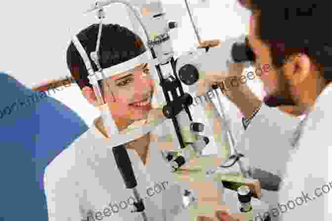
Slit Lamp Examination
A slit lamp examination is a non-invasive procedure that allows the ophthalmologist or optometrist to examine the external and internal structures of the eye in great detail. The slit lamp is a microscope that projects a thin beam of light into the eye, providing a magnified view of the cornea, iris, lens, and vitreous humor. During the examination, the patient rests their chin on a chin rest and forehead against a headband to keep their head still. The ophthalmologist or optometrist then uses the slit lamp to examine the eye from different angles, looking for signs of inflammation, infection, or other abnormalities.
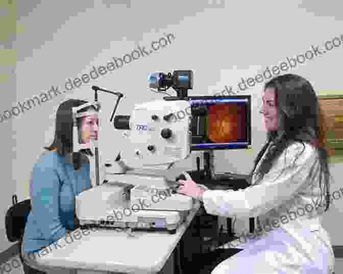
Ophthalmoscopy
Ophthalmoscopy is a diagnostic procedure used to examine the interior of the eye, including the retina, optic nerve, and blood vessels. The ophthalmoscope is a lighted instrument that allows the ophthalmologist or optometrist to view the fundus of the eye through the pupil. During the examination, the patient typically dilates their pupils with eye drops to provide a wider field of view. The ophthalmoscope is then held close to the patient's eye, and the ophthalmologist or optometrist uses various lenses to focus on different parts of the fundus. Ophthalmoscopy can detect a variety of eye conditions, such as macular degeneration, diabetic retinopathy, and glaucoma.
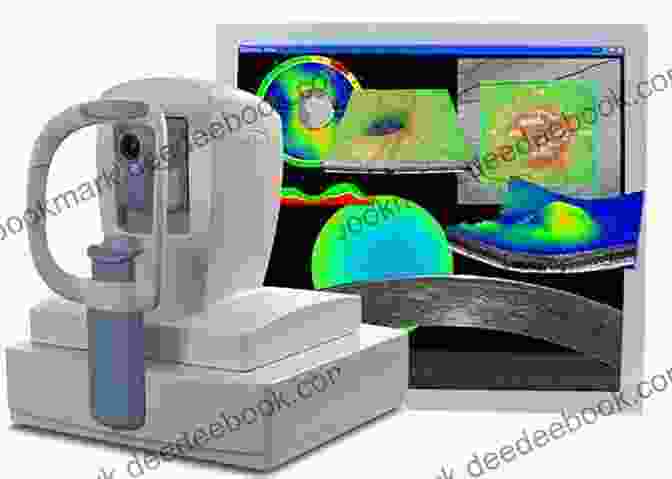
Ocular Coherence Tomography (OCT)
Optical coherence tomography (OCT) is a non-invasive imaging technique that provides cross-sectional images of the retina and other structures of the eye. OCT uses light waves to create detailed, high-resolution images that can help diagnose and monitor a wide range of eye conditions, including macular degeneration, glaucoma, and diabetic retinopathy. During the procedure, the patient rests their chin on a chin rest and forehead against a headband to keep their head still. The OCT machine then projects light waves into the eye, and the reflected light is used to create the images.
Fluorescein Angiography
Fluorescein angiography is a diagnostic procedure that uses a fluorescent dye to visualize the blood vessels of the retina and choroid. The dye is injected into a vein in the arm, and as it circulates through the body, it reaches the blood vessels of the eye. The ophthalmologist or optometrist then uses a special camera to take pictures of the retina as the dye passes through it. Fluorescein angiography can help diagnose and monitor a variety of eye conditions, such as diabetic retinopathy, macular degeneration, and retinal vein occlusion.
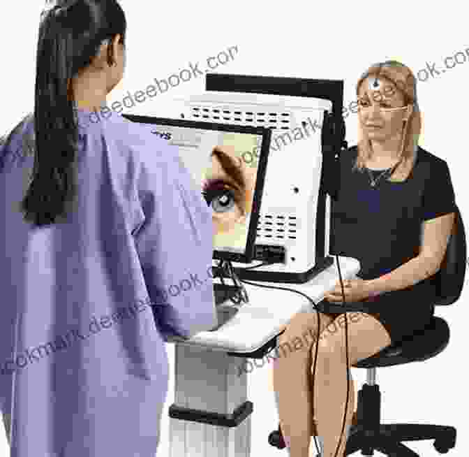
Electroretinography (ERG)
Electroretinography (ERG) is a diagnostic procedure that measures the electrical activity of the retina in response to light. During the procedure, the patient sits in a dark room and a small electrode is placed on the surface of the cornea. A series of light flashes are then projected into the eye, and the electrode records the electrical signals produced by the retina. ERG can help diagnose and monitor a variety of eye conditions, such as retinitis pigmentosa, macular degeneration, and glaucoma.
Optical Coherence Tomography Angiography (OCT-A)
Optical coherence tomography angiography (OCT-A) is a non-invasive imaging technique that combines OCT with angiography to visualize the blood vessels of the retina and choroid. OCT-A uses light waves to create detailed, high-resolution images of the blood vessels, without the need for a fluorescent dye. OCT-A can help diagnose and monitor a variety of eye conditions, such as diabetic retinopathy, macular degeneration, and retinal vein occlusion.

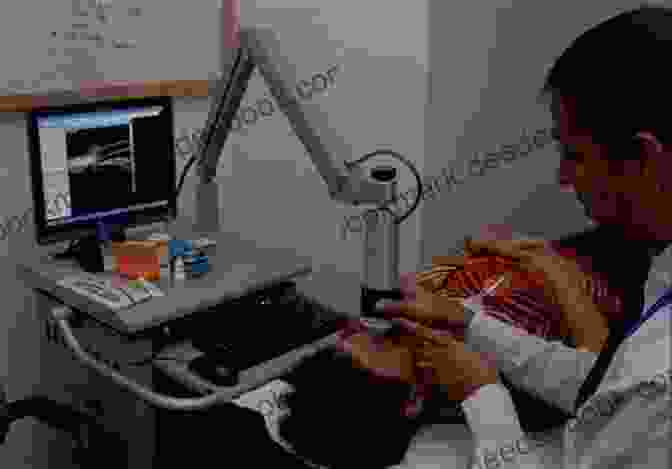
Ultrasound Biomicroscopy (UBM)
Ultrasound biomicroscopy (UBM) is a high-resolution ultrasound scanning technique that provides detailed images of the anterior segment of the eye, including the cornea, iris, and lens. UBM uses sound waves to create cross-sectional images of the eye, and it can help diagnose and monitor a variety of eye conditions, such as glaucoma, uveitis, and corneal ulcers.
Diagnostic procedures in ophthalmology play a crucial role in the diagnosis and management of eye disorders. By utilizing these advanced techniques, ophthalmologists and optometrists can accurately assess eye health, identify potential issues, and develop appropriate treatment plans to ensure optimal patient outcomes. The comprehensive overview provided in this article serves as a valuable resource for healthcare professionals seeking a deeper understanding of the various diagnostic procedures employed in ophthalmology.
4.5 out of 5
| Language | : | English |
| File size | : | 55845 KB |
| Print length | : | 462 pages |
| Screen Reader | : | Supported |
Do you want to contribute by writing guest posts on this blog?
Please contact us and send us a resume of previous articles that you have written.
 Book
Book Novel
Novel Text
Text Story
Story Reader
 Library
 Paperback
 Magazine
 Newspaper
 Paragraph
 Shelf
Shelf Preface
 Synopsis
 Annotation
 Footnote
 Scroll
 Codex
Codex Tome
Tome Autobiography
 Reference
 Encyclopedia
 Dictionary
 Thesaurus
 Librarian
 Catalog
 Card Catalog
 Archives
 Periodicals
 Study
Study Research
 Lending
 Reserve
 Academic
 Journals
 Reading Room
 Literacy
 Study Group
 Dissertation
 Reading List
 Book Club
 Franklin Colletta
 Simon Hawkins
 Ben Clanton
 Whitney Strub
 Anthony Baird
 David Schmidtz
 John Cambridge
 Chekitan S Dev
 Tarik Lebbadi
 Muhammad Rafique
 Elizabeth L Hinson Hasty
 Mary Ann Warren
 Gregory Laxer
 Richard G Lewis
 Sylvia Ellis
 Michael Bond
 Nicola Reeder
 Ant Mcpartlin
 Baz Kershaw
 Jonathan Beam
Light bulbAdvertise smarter! Our strategic ad space ensures maximum exposure. Reserve your spot today!

 Winston HayesAn Open Letter to the Women Who Will Run the World: Our Future Depends on You


 Caleb CarterUnveiling the Magic of Dog Training with JK Brandon: A Comprehensive Guide to...
 Damon HayesFollow ·5.5k
 Elmer PowellFollow ·17.7k
 Franklin BellFollow ·10.3k
 Lord ByronFollow ·8.4k
 Fredrick CoxFollow ·16.8k
 Eugene PowellFollow ·12k
 Thomas PynchonFollow ·12.1k
 DeShawn PowellFollow ·14.5k

 Oscar Wilde
Don't Stop Thinking About the Music: Exploring the Power...
Music is an...

 Floyd Richardson
Snowman Story Problems Math With Santa And Friends
It's a cold winter day, and...

 W. Somerset Maugham
What Every Classroom Teacher Needs To Know: A...
Teaching is a challenging...

 Edgar Cox
Tall Tales But True: A Lifetime of Motorcycling...
I've been riding motorcycles for over 50...

 Chinua Achebe
Buni: Happiness Is a State of Mind
Buni is a beautiful...

 Herman Melville
The Arts and Crafts of Older Spain: Embodying the Essence...
In the heart of the Iberian...
4.5 out of 5
| Language | : | English |
| File size | : | 55845 KB |
| Print length | : | 462 pages |
| Screen Reader | : | Supported |
